Overall, I thought that the 20 time presentations were really interesting to listen to. I noticed how not one presentation was the same, each covering their own unique topic. I thought it was also interesting how some topics were briefly mentioned in others, but the process and end result was completely different. It was really cool to hear how other people's projects went as a whole, and knowing that you were not the only one who struggled.
Tuesday, June 6, 2017
20 Time Final Post
I finally did my 20 time TED talk with Justine on Thursday, June 1. I thought that it went a lot better than I thought it would go. I thought that we did a good job highlighting all of our key points that we wanted to cover. Also, I thought that we did a good job reflecting on our journey as a whole. However, after watching myself present, I have realized that I look as uncomfortable presenting as I feel. I kept fidgeting on my crutches and looking over at Justine. Instead of doing this, I should have just relaxed more and looked at the audience when I was not talking. I would have given myself an A because while I was not the best presenter, I was able to explain all the key points I wanted to say and we covered the requirements. I have grown from this experience because it was another opportunity to give a presentation in front of others. No matter how many times I practice, I am always super nervous to present. This was just another chance for me to practice my public speaking and assess myself afterwards, which is not something that I can normally do.
Thursday, May 25, 2017
20 Time Reflection
When I first started this project, I was very lost. I had trouble grasping the idea of 20 time and what I could even do for the project. I ended up spending a solid chunk of my time trying to figure out what I was going to do. Eventually, I talked to Justine and we decided that we should do something together since we both did not know what we were doing for our respective projects. Just like before, we both struggled in figuring out what we should do. We started listing out different topics that we just enjoyed and would possibly want to look into. While the topics we discussed were very interesting, they were not the easiest ones to use to develop a final product with. We were cutting it down to the wire for our topic, and on the day that we had to turn our project proposal in, I had read an article on California’s water management. It talked about the usage of groundwater and how that is pumping the water out of the Earth, drying it out. After reading this article, I was fascinated with the topic and proposed it to Justine. We both agreed to it since it is relevant to us and it seemed like a good topic to choose for our project.
As we began to execute our plan, things did not go the way that we wanted it to. We overestimated our abilities to create a product that would be informative and interesting. Initially, we had this great plan to make a website that would inform people about our cause and how to help better manage water. Additionally, we had planned to include a documentary that could visually show an audience what exactly is going on, and what the public can do. Unfortunately, all of this did not go as planned so we had to completely scratch that idea. Instead, we have decided to create an infographic to present different information that we have found. This is what we have made.
This has been a really frustrating and stressful process for me. I do not think that I have had to change directions for a project so drastically before. It is definitely a different process, whether it be good or bad is still to be determined. I feel like through the constant changing of ideas has really forced me to be able to adapt to anything fast so that progress continues to be made. I have learned how to think on my feet and find a solution when things do not go as planned. This happened a lot to us unfortunately, but it did help me gain this skill. If I ever were to do this same project again, I would make sure to avoid electronics for the final product. Instead, I would look to do something that I could maybe test out or just have more concrete things.
I plan on taking all of the skills and information that I have learned from this project and apply them to my decisions in the future. I am definitely going to be more conscious about water and how I manage it from now on. Also, I plan on trying to include some of these tips I have learned at my own home.
Based off of everything that we had to deal with and what we were able to accomplish, I would give myself an A on this project. I made sure to always keep up with the blog posts with Justine, where we talked about our progress for each of the required blog posts: 1, 2, 3, and 4. Also, I would comment on other people’s posts and give my opinion and advice to others on how they could improve their project. I would always work to my best ability during class to try to improve our project and come up with creative ideas.
Ted Talk Outline:
1. Why we chose this topic
1. Why we chose this topic
2. Specific facts regarding this topic
3. Show the infographics and elaborate more
4. How everybody can help manage water
Monday, May 15, 2017
Unit 8 Reflection
This unit was primarily about the muscular system and the movements that we make. We began this unit by learning about the different synovial joint movements. Some movements that we went over included flexion, which decreases the angle between articulating bones, and extension, which increase the angle between articulating bones. We also learned the six synovial joints: plantar, hinge, pivot, condyloid, saddle, and ball and socket. I studied the ACL in the knee in particular when completing build a better joint project. Plantar joints are flat and slightly curved joints that are commonly found in feet. Hinge joints are convex surfaces where one bone fits into the concave of another. Pivot joints are rounded or pointed surface of one bone that articulates with a ring formed partly by a ligament. Condyloid joints are convex oval-shaped projections of one bone that fit into the concave oval-shaped depression of another bone. A saddle joint is when the articular surface of one bone is saddle-shaped, while the articular surface of the other bone fits into the saddle like a rider on a horse. A ball and socket joint is when a ball like surface of one joint fits into the cup like depression of another bone.
We also learned about the different properties of muscles and their functions. Muscles have four main functions: movement of bones and fluid, body posture and position, stabilizing joints, and heat generation. The properties of muscles are contractility, which is the ability to shorten when stimulated; excitability, which is the ability to receive and respond to stimuli; extensibility, which is the ability to be stretched; and elasticity, which is the ability to recoil to resting length. All muscles have at least one origin, which is the immovable end of a muscle, and one insertion, which is the end of the muscle that moves towards the origin. Muscles are classified by their function. The prime mover causes a desired action, while the anatagonist relaxes when the prime mover contracts. The synergist helps the prime mover by reducing unnecessary movements. Fixators help stabilize the origin of the prime mover. We also went over some commonly known muscles such as the biceps brachii, which flexes the elbow joint, and the triceps brachii, which extends the elbow joint. We were able to examine many of these muscles for ourselves when we did our chicken dissection.

Each muscle fiber is composed of myofibrils and sarcomeres. Muscles contract with the sliding filament theory. To signal a muscle contraction, nerves send impulses to the muscle fibers, resulting in Ca+ being released. The Ca+ binds to the receptors on the fiber, causing the myosin-binding sites to be open. The opening of these binding sites allows the myosin to slide the muscle into contraction, with the help of ATP. There are three different types of muscle fibers: Slow twitch, fast twitch a, and fast twitch b. Slow twitch fibers are generally found in long distance runners since they contract at a slower rate and can last longer. They are dependent on blood for oxygen and nutrients so they are red. Fast twitch a fibers contract at a much faster rate and are relatively fatigue resistant due to their relatively high blood flow capacity. On the other hand, fast twitch b fibers fatigue quickly because of their low blood flow capacity. When an individual exercises, their muscles can either go through hypertrophy, where cells increase in size/volume due to myofibrils, or hyperplasia, where cells remain the same size, but increase in number.

Muscle contractions are classified into four different groups. In concentric contractions, the muscle is actively shortening, while in eccentric contractions, the muscle is actively lengthening. Isometric contractions are when the muscle is actively held at a fixed length. An example of an isometric contraction is holding a plank. Lastly, passive stretching is when the muscle passively lengthens.
I want to learn more about what happens during a muscle cramp or twitch. I know that cramps have something to do with calcium, so maybe they occur due to low levels of calcium during a muscle contraction, causing the whole muscle to get stuck in a contraction. I have grown as a student through the different activities and labs that we do in class. For example, through the chicken dissection, I was able to identify different muscles on an actual chicken that I eat on a regular basis. It really showed me the complexities of something that seems so simple. I have been doing a good job in one half on my new years goal in that I have really been relating what I am learning to my everyday life. This whole unit has helped me understand my own body better and the reasons behind some of my physical therapy exercises. I am also doing pretty well on the eating healthier part of my goal. I think that will be very important for me as I continue recovering from my knee surgery. And of course I am still unable to move around well so the exercising part of my goal will probably have to be taken out.
Monday, May 8, 2017
What happens when you stretch
This article discussed the importance of stretching and how the muscles are affected when they are stretched. Stretching is vital to the health of muscles because when the muscles stretch, the muscles lengthen. The two types of muscle fibers are intrafusal muscle fibers and extrafusal muscle fibers. Intrafusal muscle fibers lie parallel the the extrafusal muscle fibers, which are the ones that contain myofibrils. Proprioceptors are nerve endings that relay information about the muscles to the central nervous system. They are used in stretching to send messages about the muscles lengthening too much or too suddenly. The stretch reflex is the reflex where the muscle attempts to resist muscle lengthening by contracting. Over time this reflex can be slowly reduced by a continuation of stretching it so that the signal is ignored by the body. Dynamic stretching relates to movements that only have sudden increases in muscle lengths, while static stretching involves long lengths of the muscle being stretched. Nuclear chain fibers are responsible for static stretching, while nuclear bag fibers are responsible for the dynamic portion. When one muscle contracts, it causes another muscle to relax and lengthen. This is known as reciprocal inhibition.
"The proprioceptors detect any changes in physical displacement (movement or position) and any changes in tension, or force, within the body."
I found this quote really interesting because I never knew about proprioceptors before. I had always assumed that there were just average neurons and nerves in the body that sense these things. Do malfunctions of proprioceptors contribute to muscle cramps or twitches?
"This triggers the stretch reflex (also called the myotatic reflex) which attempts to resist the change in muscle length by causing the stretched muscle to contract. The more sudden the change in muscle length, the stronger the muscle contractions will be"
I found this interesting as well because I have experienced this myself many times. This reflex happens when I try to stretch, showing me that I am not flexible and have to go slower. It helps me realize my limits.
"When this tension exceeds a certain threshold, it triggers the lengthening reaction which inhibits the muscles from contracting and causes them to relax"
I still find this phenomenon really interesting. I also experienced this first hand with the hamstring stretch and saw how much farther my leg went after flexing my quadricep. How can the line between healthy and excessive be drawn for stretching?
Designing a More Effective Joint: The Knee
In this project I researched the anterior cruciate ligament to figure out how to make it better. The knee is a complex joint of the human body that can easily be hurt. The anterior cruciate ligament (ACL) is one of the most common serious knee injuries. It can easily be hurt through sports and the recovery time is a long one. I researched key ways the ACL is damaged and came to the conclusion that it should be modified by increasing the thickness of it, adding more muscle tissue, and adding more motor neurons. These theoretical additions will decrease the chance of injury and improve the joint. In reality, the knee cannot be redesigned, so ways to protect this ligament include keeping the thigh muscle strong, warming up before exercise, and maintaining flexibility.
 |
| My knee 2 days after the injury occurred |
My new redesign of the ACL could theoretically reduce likelihood of injury in the first place. In the new design of the knee, the ACL, which is usually about 10 mm thick on average, would be 2 mm thicker right and left, which a total addition of 4 mm of ligament. The addition of this extra ligament could hopefully reduce the likelihood of the ligament tearing and over stretching. The new knee would also have a larger muscle mass. The muscle of the hamstring in particular would be increased by about 6 cm in diameter. This would allow the hamstring to naturally be stronger and help allow it to absorb more of the shock placed on the knee. The quadricep would also be increased by about 4 cm in diameter. Like the hamstring, the quadricep would help protect the ACL from injury by being able to absorb more shock and have more strength to make safe movements. The hamstring is increased more than the quadricep because many people naturally have weaker hamstrings, so more muscle should be added. I would also increase the number of motor neurons so that individuals would have a quicker reaction time if they were in a situation that could lead to injury. These neurons would allow an individual to have a better sense of their own body, reducing the chance of injury.
This new diagram of the ACL displays how it is approximately 4 mm larger than it was originally. The thinness of the original ligament plays a role in how it tears and stretches easily. The new thickness allows the ligament to be tougher.
 |
| Before |
 |
| After |
This diagram displays the difference in structure in the additional muscles added to the improved knee. The increase of the muscle mass makes the leg stronger than it was before so that the ACL will not be as traumatized in different situations.
 |
| Before |
 |
| After |
I arrived at this design through my prior knowledge of the ACL and additional research that I did. Ideally, I would have decreased the width of women’s hips, but that would simply not work out since they are that way so that they can give birth to children. A problem that I ran into was trying to figure out reasonable sizes of the muscles and ligaments. I could not find any source that explicitly stated the size of the hamstring or quadriceps muscle, so I had to use common sense. A problem with increasing the width of the ACL is that it may reduce the mobility of the knee if the thickness reduced movement; every part of the body is made how it is for a reason. The increase in motor neurons may also be a problem if it creates too much information for the brain to process, which could possibly lead to seizures if it gets too much. This relates to what we have learned in class because we have learned about what ligaments are and how muscles always have another one to work inversely with it. The increase in size of the hamstring and quadriceps is an example since when the hamstring flexes, the quadricep relaxes. Because we are unable to actually redesign different joints, different things can be done to prevent this injury. One way to prevent injury is to keep the thigh muscles strong. The strengthening of these muscles allows legs to stay strong and add extra protection to the ACL. Another way to stay healthy is to warm up before doing strenuous exercise. This will allow muscles to get moving so that they are able to support sudden movements. Maintaining flexibility will also help so that an individual’s body is able to have full range of motion without straining it.
Works Cited
"Anterior Cruciate Ligament (ACL) Injuries - Topic Overview." WebMD. WebMD, n.d. Web. 09 May 2017.
"Anterior Cruciate Ligament (ACL) Injuries." Anterior Cruciate Ligament (ACL) Injuries-OrthoInfo - AAOS. N.p., 01 Mar. 2014. Web. 09 May 2017.
Hindawi. "Adequacy of Semitendinosus Tendon Alone for Anterior Cruciate Ligament Reconstruction Graft and Prediction of Hamstring Graft Size by Evaluating Simple Anthropometric Parameters." Anatomy Research International. Hindawi Publishing Corporation, 29 July 2012. Web. 09 May 2017.
Horwitz, D.C. Steven. "ACL Injuries: Female Athletes At Increased Risk." MomsTeam. N.p., n.d. Web. 09 May 2017.
"KneeAnatomy Side." Wikipedia Commons. Wikipedia Commons, n.d. Web. 8 May 2017.
Mysid. "Knee Diagram." File:Knee Diagram.svg - Wikimedia Commons. Wikipedia Commons, n.d. Web. 09 May 2017.
Tortora, Gerard J., and Bryan Derrickson. Introduction to the Human Body: The Essentials of Anatomy and Physiology. New York: Wiley, 2007. Print.
"What Are Knee Ligament Injuries?" WebMD. WebMD, n.d. Web. 09 May 2017.Wednesday, May 3, 2017
Chicken Dissection Lab Analysis
 |
We began the dissection by opening up the chicken's chest and identifying the pectoralis major, pectoralis minor, and the sternum. The pectoralis major is in charge of pulling the wing ventrally to power the flapping of wings for flight. This muscle works with the pectoralis minor, which lifts the wing dorsally. The sternum is in the middle of the bird, connecting the muscles on both sides of the body can work together in unison to enable the act of flight. The sternum is very long and curved, which gives support to the large pectoral muscles and allows them to be the size that they are. The tendons connect the muscles to the sternum, acting as the glue to make everything work.
 |
| Anterior view of chicken Blue: Sternum- connects to pectorals Red: Pectoralis Minor-triangularly shaped, pulls wing dorsally White: Pectoralis Major- triangularly shaped, pulls wing ventrally |
 |
| Green: Deltoid- triangular, raises upper arm or wing Black: Bicep brachii- has two origins, flexes wing or arm Yellow: Tricep humeralis- has three origins, flexes wing or arm |
We also dissected the chicken wing, where we were able to observe the different roles that the tendon's origin and insertion play. The origin of a tendon is the immovable end of the muscle. The point of insertion is the movable end of the muscle. In the wing, the triceps humeralis, biceps brachii, and deltoids came into play. The biceps contract the muscle, which brings the point of insertion closer to the origin. The tendons connect the muscles together at the joints, allowing them to flex and straighten. The action of these muscles allows for the flapping of wings.
 |
| Yellow: Flexor carpi ulnaris- The largest muscle on the posterior side of the lower wing away from the ulna. It flexes the hand. |
 |
| Blue: Brachioradialis- largest muscle on the superior side of the lower wing closest to the alua. It pulls the hand back |
Humans and chicken have similar, yet different muscles. Just like a chicken, humans have pectorals; however, the pectorals of humans are not as large as they are on a chicken. Birds, unlike humans, require strong pectoral muscles so that they can flap their wings at a constant rate. On the other hand, humans do not raise their arms that much in their day-to-day activities. Both humans and chicken have a trapezius, which is located on the upper back. In the chicken, the muscle runs from the backbone to the shoulder, and the shoulder to the back. On the other hand, in humans, the trapezius is split into two separate parts. In humans, the trapezius shrugs the shoulders and pulls the shoulders back. On the other hand, because the muscle is one main part in the chicken, I believe that the trapezius primarily helps pull up the arms.
 |
| Posterior View of Chicken Green: Trapezius- trapezoidal, pull shoulders back Yellow: Latissimus dorsi- help extend or pull the wing or arm |
Another similar muscle between humans and chicken is the peroneus longus. The main difference between these muscles in humans and chickens is the relative size of the muscle in comparison to chicken. Chicken calves are completely round, unlike a human's. While I am unsure why chicken calves are so large, it may have to do with the fact that they walk in a squatted-like position and need the muscle, or they are bred that way so that consumers get more meat per bone.
 |
| Calf Blue: Peroneus longus- longest muscle in calf, extends the foot Yellow: Gastrocnemius- extends foot and flexes the lower leg Green: Tibialis anterior- flexes the foot |
 |
| Anterior view of thigh Yellow: Quadriceps femoris- four origins, flexes thigh and extends the lower leg Blue: Sartorius- flexes thigh and allows crossing of the legs Green: Semitendinosus- extends the thigh |
 |
| Posterior view of thigh White: Biceps femoris- has two origins, flexes the leg Black: Iliotibialsi- extends the thigh and flexes the leg Red: Semimembranosus- extends the thigh |
Sunday, April 16, 2017
Unit 7 Reflection
In this unit we learned about the skeletal system and different disorders that can occur. Bones are divided into the axial system, which is composed of the skull, spine, and ribcage, and the appendicular skeleton, which is composed of all the other bones. With in the two different divisions, bones are classified in different ways. There are short bones, which are shorter than they are wide; long bones, which are longer than they are wide; flat bones, which are thin and flattened; and irregular bones, which have an irregular shape and do not fit into any of the other categories. In our Owl Pellet Lab we were able to dissect an actual owl pellet and identify the organism that it had eaten based off of what the bones looked like. Two types of bone tissue are compact and spongy bones. Compact bones are strong, while spongy bones are full of small spaces and are more flexible. There are different types of bone cells within the bones themselves. Osteocytes are mature bone cells. Osteoblasts are bone-forming cells, while osteoclasts are bone destroying cells. The balance of all of these cells allows the bones to grow and stay healthy and strong. Bones receive nutrients from blood vessels that run through them. The Haversian canal contains at least one blood capillary which is the source of nutrients and means of waste disposal.

There are many different disorders that can occur in the skeletal system. Arthritis is the inflammation of the joints, which is caused by age, infection of the joints, and trauma from the joints. Unlike what many people believe, cracking one's knuckles does not lead to arthritis. Osteoporosis is the condition in which bones have lost minerals and the holes become too large, allowing the bones to become brittle. Scoliosis is when there is an abnormal curvature of the spine. This is more common in girls than boys. Kyphosis is when the spine may develop a hump, affecting people of all ages. Lordosis is the excessive curvature of the lumbar spine and forward pelvic tilt. This is common in gymnasts and dancers. Rickets is the softening and weakening of bones, which is common in children with vitamin D deficiency.
Ossification is the process by which a bone forms. This process usually continues until the individual is 25 years of age. Epiphyseal Disks are responsible for longitudinal growth. The calcium that is stored inside bones helps maintain the blood pH levels and body fluids, heart function, muscle contractions, nerve function, and blood clotting. The osteoblasts in the bones produce osteocalcin, which is a calcium binding cell. The osteoclasts secrete enzymes that help function in reabsorption. The hormone Parathyroid hormone increases the number of active osteoclasts, which will increase the amount of calcium. Calcitonin is secreted to lower the level of calcium by inhibiting the osteoclasts. While calcium is necessary for bones, it cannot be absorbed without the help of vitamin D. An excess amount of sodium and protein can lead to an increase of calcium excretion because of the build up of amino acids in the blood. Overall, it is vital to keep your bones healthy through exercise and a well balanced diet. Bone fractures may occur if a more force is put on a bone than it can hold. There are different types of bone fractures. A complete fracture is when the bone is broken completely through. An incomplete or greenstick fracture is when the fracture does not extend through the bone. A closed fracture is when the bone does not tear through the skin. A compound fracture is when skin is pierced. A comminuted fracture is when the bone is fragmented into three or more pieces. An oblique fracture occurs when the bone breaks at an angle. Once the bone breaks, it bleeding and inflammation occurs, producing a blood clot. The clot then softens after about 48 hours, producing a procallus. Then, fibroblasts arrive and secrete dense connective tissue that replaces the procallus. Chrondroblasts and osteoblasts are next to arrive. After the first week cartilage and bone appear throughout the site. An osseous callus acts as an internal splint for the site. Once the osseous callus has undergone remodeling to restore the bone, it is healed.

A joint is the point of connection between two bones or elements. There are three classifications of levers for joints: First-class lever, which have a fulcrum in the middle between effort and resistance; second-class lever, which has resistance between fulcrum and effort; and third-class lever, which have effort between resistance and fulcrum. In terms of movement, joints can be synarthroses, immovable; amphiarthroses, or slightly movable; diarthroses, or freely movable. Structurally, bones can be fibrous, which are generally immovable; cartilagenous, which are immovable or slightly movable; or synovial, which are freely movable. A joint that is fibrous and synarthroses is an immovable fibrous joint. An example of this would be the skull. A fibrous and amphiarthoses joint would be bones that are only connected to ligaments. a cartilagenous and synarthroses joint would be bones that are joined by the hyaline cartilage. These are generally immovable. A cartilagenous and amphiarthoses joint is bound by the fibrocartilage and is slightly movable. a synovial and diarthroses joint is a joint between two tones separated by a space called a joint cavity. A bursar is a small fluid-filled sac that is linked by synovial membrane with inner capillary layer of slimy fluid. This provides a cushion between the bone and tendons and reduces the friction.

I want to learn more about topics that are relevant to me. For example, I want to know about what a stress fracture would be. Also I'm curious about common injuries in the joints, such as torn ACLs menisci. I still struggle in distinguishing between the different joints and the combinations of them together. Also, I'm curious about how the different ligaments in the knee helps keep it stable.
I think I have grown throughout this whole unit as a student. During the Owl Pellet Lab, I learned the importance of patience and considering many different options because the bones were scrambled and there could have been more than one organism in the pellet. I also have more of an appreciation for what we are learning because I hurt my knee and we learned about the different joints in this unit. Also, the fractures section relates to me because I fractured one of my fingers when I was in elementary school. The swelling of the finger makes much more sense now since before I never really knew the reason behind it. I can make sure to keep my bones healthy by making sure I go outside enough and get vitamin D so that the calcium that I take in can be absorbed into bones. My goal of running is going to be difficult for the next 6ish months since I am unable to run with my bad knee. I could just work on strengthening my legs instead.

There are many different disorders that can occur in the skeletal system. Arthritis is the inflammation of the joints, which is caused by age, infection of the joints, and trauma from the joints. Unlike what many people believe, cracking one's knuckles does not lead to arthritis. Osteoporosis is the condition in which bones have lost minerals and the holes become too large, allowing the bones to become brittle. Scoliosis is when there is an abnormal curvature of the spine. This is more common in girls than boys. Kyphosis is when the spine may develop a hump, affecting people of all ages. Lordosis is the excessive curvature of the lumbar spine and forward pelvic tilt. This is common in gymnasts and dancers. Rickets is the softening and weakening of bones, which is common in children with vitamin D deficiency.
Ossification is the process by which a bone forms. This process usually continues until the individual is 25 years of age. Epiphyseal Disks are responsible for longitudinal growth. The calcium that is stored inside bones helps maintain the blood pH levels and body fluids, heart function, muscle contractions, nerve function, and blood clotting. The osteoblasts in the bones produce osteocalcin, which is a calcium binding cell. The osteoclasts secrete enzymes that help function in reabsorption. The hormone Parathyroid hormone increases the number of active osteoclasts, which will increase the amount of calcium. Calcitonin is secreted to lower the level of calcium by inhibiting the osteoclasts. While calcium is necessary for bones, it cannot be absorbed without the help of vitamin D. An excess amount of sodium and protein can lead to an increase of calcium excretion because of the build up of amino acids in the blood. Overall, it is vital to keep your bones healthy through exercise and a well balanced diet. Bone fractures may occur if a more force is put on a bone than it can hold. There are different types of bone fractures. A complete fracture is when the bone is broken completely through. An incomplete or greenstick fracture is when the fracture does not extend through the bone. A closed fracture is when the bone does not tear through the skin. A compound fracture is when skin is pierced. A comminuted fracture is when the bone is fragmented into three or more pieces. An oblique fracture occurs when the bone breaks at an angle. Once the bone breaks, it bleeding and inflammation occurs, producing a blood clot. The clot then softens after about 48 hours, producing a procallus. Then, fibroblasts arrive and secrete dense connective tissue that replaces the procallus. Chrondroblasts and osteoblasts are next to arrive. After the first week cartilage and bone appear throughout the site. An osseous callus acts as an internal splint for the site. Once the osseous callus has undergone remodeling to restore the bone, it is healed.
A joint is the point of connection between two bones or elements. There are three classifications of levers for joints: First-class lever, which have a fulcrum in the middle between effort and resistance; second-class lever, which has resistance between fulcrum and effort; and third-class lever, which have effort between resistance and fulcrum. In terms of movement, joints can be synarthroses, immovable; amphiarthroses, or slightly movable; diarthroses, or freely movable. Structurally, bones can be fibrous, which are generally immovable; cartilagenous, which are immovable or slightly movable; or synovial, which are freely movable. A joint that is fibrous and synarthroses is an immovable fibrous joint. An example of this would be the skull. A fibrous and amphiarthoses joint would be bones that are only connected to ligaments. a cartilagenous and synarthroses joint would be bones that are joined by the hyaline cartilage. These are generally immovable. A cartilagenous and amphiarthoses joint is bound by the fibrocartilage and is slightly movable. a synovial and diarthroses joint is a joint between two tones separated by a space called a joint cavity. A bursar is a small fluid-filled sac that is linked by synovial membrane with inner capillary layer of slimy fluid. This provides a cushion between the bone and tendons and reduces the friction.
I want to learn more about topics that are relevant to me. For example, I want to know about what a stress fracture would be. Also I'm curious about common injuries in the joints, such as torn ACLs menisci. I still struggle in distinguishing between the different joints and the combinations of them together. Also, I'm curious about how the different ligaments in the knee helps keep it stable.
I think I have grown throughout this whole unit as a student. During the Owl Pellet Lab, I learned the importance of patience and considering many different options because the bones were scrambled and there could have been more than one organism in the pellet. I also have more of an appreciation for what we are learning because I hurt my knee and we learned about the different joints in this unit. Also, the fractures section relates to me because I fractured one of my fingers when I was in elementary school. The swelling of the finger makes much more sense now since before I never really knew the reason behind it. I can make sure to keep my bones healthy by making sure I go outside enough and get vitamin D so that the calcium that I take in can be absorbed into bones. My goal of running is going to be difficult for the next 6ish months since I am unable to run with my bad knee. I could just work on strengthening my legs instead.
Friday, March 31, 2017
Owl Pellet Lab Analysis
 In this lab we analyzed the contents of an owl pellet. The goal of doing this was to determine the organism that the owl had eaten by analyzing the different bone structures of the animal. By inspecting these animal bones the type of animal it was can be determined by looking at key features of different parts of the bone. In this lab, we were given a chart that displayed the different types of animals that it could be and pictures of the unique bone structures. Some key features that we looked for in our animal included the skull, pelvis, tibia, and fibula. Unfortunately, the specimen that we obtained did not contain a skull so different means of identifying the animal had to be taken. Instead, we primarily focused on identifying the pelvis, tibia, and fibula.
After putting together all the bones that we found, we determined that the animal that we obtained was a vole. Although the data was somewhat inconclusive, some key features from the bones pointed to the animal being a vole. We were able to come to this conclusion because the some key features in our specimen matched the features in the chart that was given. For example, the pelvis of our animal matched the most closely to the pelvis of a vole. The triangular shape of the pelvis matched the picture of it on the chart that we got. Also, the superior side of the bones matched the thickness of the picture of the vole's pelvis. The gap that the bones make when they meet also was similar to the chart since it was much smaller than that of a shrew's. Also, the tibia and fibula were very similar to the vole's. The curve of the fibula when it bend over to the tibia was similar in that it was bent at about a 90 degree angle. This key feature distinguished it from that of a shrew's because the shrew's fibula bent at more of a 120 degree angle.


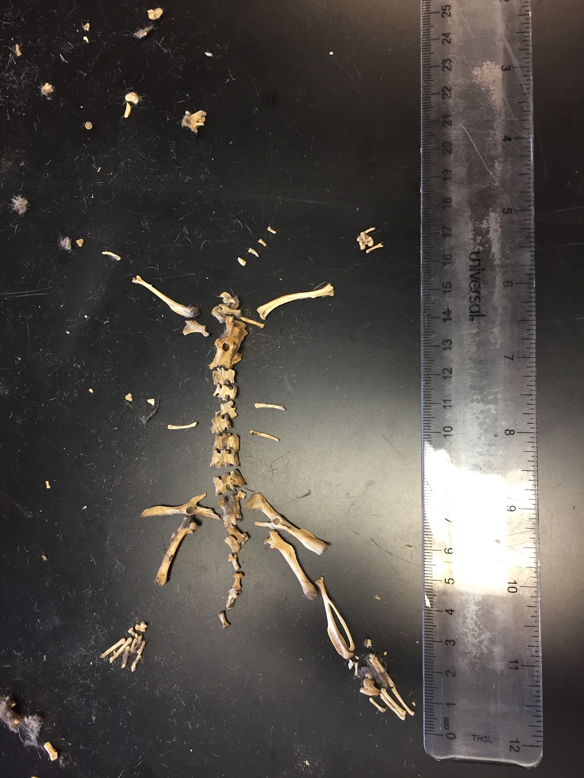
The fibula and tibia were similar to the ones on the human body because they are both connected and located in the lower leg. In both the human and vole, one bone was larger than the other; however, in the vole, the fibula was larger, while in a human the tibia is much larger. Also, the gap between the two bones on the vole was much larger and circular than it is in a human. This displays how these organisms have very similar features, but are unique in their own ways.


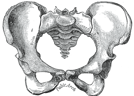
Another structure that was similar to a human's was the pelvis. Both pelvises have a gap, where the bones form loops. However, the pelvis of the vole is more triangularly shaped than the human's. The human pelvis is much wider and contains another large gap in it.

The spine of a vole was remarkably similar to the spine of a human. Both of the spines had individual pieces that connected together. Both spines appeared to have jagged edges, but the individual pieces of a human spine are much more round. The voles' spine is very jagged all around.


Nervous System Extra Credit
Your Brain on Food: How Chemicals Control Your Thoughts and Feelings By: Gary L. Wenk

In book titled Your Brain on Food, which is written by Gary L. Wenk, talks about how the many different neurotransmitters and food in our everyday diet affect our brain. Throughout this whole book, Wenk expands on the concept that he explains by bringing up different instances that he himself has experienced, or past examples in history. He is successful in creating a compelling book that is both engaging and informative. The main point that Wenk argues in his writing is that the brain is an advanced organ in our body that can constantly become affected by our many foods in our diet. Because of this people need to eat less food since food in volumes because too much of anything never has positive health effects in our body for the long run, which can cause long lasting effects.
In the book, Wenk brings up lots of different drugs and neurotransmitters that each affect the brain in its own unique way. Throughout the whole book he explains how the drugs affect the brain and how nowadays scientists are able to change the chemical components of drugs to enable them to be more effective by making them more lipid soluble, allowing them to pass through the blood-brain barrier with much more ease. This scientific breakthrough allows drugs to have a much more potent effect on the body than the original drug would: “morphine, for example, became far more lipid-soluble and far more euphorigenic (i.e., pleasure-inducing) when scientists added two acetyl groups to it to produce heroin at the turn of the 19th century. Much later, amphetamine was similarly modified to make it more euphorigenic and therefore more addicting” (Wenk 58). This displays how this minor alteration of adding methyl groups completely changes the chemical structure of the drug, allowing it to become more addicting and have a greater effect on the user. This exhibits how people need to definitely be weary about what they take and the amount that they use because many drugs today are able to have a greater impact in the brain because of their new lipid solubility. Another example of this changing structure is the formation of ecstasy, which can have many adverse effects. Wenk describes how some of them include, “a dramatic rise in body temperature, or hyperthermia”(59). This depicts the serious consequences of taking such a potent drug such as ecstasy; Wenk warns readers to take caution in these warnings. Another drug, cocaine, is also described in this book. The author again warns readers about the negative side effects of this drug in large quantities through his in depth explanations and descriptions of what could happen: “excessive, long-term, intravenous use of cocaine tends to produce especially severe rebound phenomena, including psychotic behaviors together with delusions of grandeur and hallucinations” (72). This caution supports Wenk’s ongoing message to readers about staying smart about choices they make and to always take things in small quantities. His warnings allow readers to understand the dangerous possibilities of these drugs. Wenk later goes on in the book to talk about an everyday drug, caffeine, but more specifically coffee. While in small amounts it can have its benefits, coffee is able to affect people in different ways. A person that is extremely tired and drowsy will feel more alert after a cup of coffee, while a person that is well rested would not get that same effect. He explains how the coffee itself can have positive health benefits such as being, “a rich source of antioxidants caffeic, chlorogenic, coumaric, ferulic, and sinapic acids and silverskin… coffee drinking has been associated with a significantly lowered risk of developing Parkinson’s disease… moderate coffee-drinking of about two to three cups each day might reduce your chance of developing Alzheimer’s disease” (125-126). This displays how coffee does indeed have its own benefits, but in large quantities it is not good, as revealed through one of his students, where the student, “finished off the entire container of 32 packets [of instant coffee right out of the box]... three days later, he stopped having explosive diarrhea and finally fell asleep completely exhausted” (127). This is a strong example of how anything in large quantities is not good for you, which is what Wenk is able to argue for throughout this whole book.
This book relates to what we have learned in class because it is all about the brain and some of the nervous system. This book explains how the blood brain barrier, which we have learned about in class, only allows lipid soluble hormones to cross, so water soluble hormones are unable to get through. This goes further into what we learned in our brain unit because we learned about all three of these concepts, but never exactly went into detail about what is able to pass through the blood brain barrier. This book also discusses action potentials, which is how neurons pass a signal down to each other. It went into detail about how that signal can become disrupted through different drugs. One main theme that was prevalent in this book that is similar to what we learned in class is the idea of simply eating healthy and getting a moderate amount of exercise. Wenk explains in his book that our body’s daily routine of metabolizing food is actually what causes our body to grow old. Because of this, people need to take care of themselves to last longer by exercising and eating the right foods. If I had the opportunity to ask this author two questions one would be how did all of these people in the past know so much about drugs? Were some lethal plants just common knowledge back in Shakespeare’s time? The other question would be why do some people get the “munchies” (102) after taking marijuana?
I think that the credibility of this author is very high; however, the credibility of this material at this time may not be that great. I am sure that back when it was first released it was extremely credible, but that was seven years ago; lots of things can change in seven years. This author is a professor at The Ohio State University in the Departments of Psychology and Neuroscience and Molecular Virology, Immunology and Medical Genetics. He also has a PhD. I believe that the ideas explained in this book are very realistic. For example, Wenk explains how the placebo effect can cause many people to believe that they feel better, when in actuality they made themselves better by believing that some medicine healed them. I believe this is very real because I myself have experienced it and he explained some experiments that were done to test it out. He also explains how drugs that are more lipid soluble do not make your brain smarter, it simply works faster. He used a good analogy with a computer, explaining how newer computers are not any more smart than they were before; they just work much faster. This is how drugs work as well. It allows people to think quicker, which can sometimes seem as though they are smarter when they really are not. He then went on to explain that people that tap their fingers fast while thinking naturally think faster. I liked this idea because it made a lot of sense to me once he explained it. Also, I found it interesting that finger tapping correlates with the speed of a person’s thought process. The implications of this work are to inform the public about different drugs and how they affect the body. Wenk gets this and the message to take caution and use small amounts of everything across in his writing. This is not theoretical because Wenk describes many different experiments that have been performed to test these facts. This benefits individuals because it allows them to understand what is actually happening to them when they take a certain type of drug, and what some side-effects could be.

Thursday, March 16, 2017
Unit 6 Reflection
This unit was all about the brain and its behavior. In this we learned about the brain and the many different structures and the functions of them as well. The brain is composed of the forebrain, midbrain, and hind brain. It is also has many other parts. The cerebrum is located in the area where the forebrain is. It is sectioned into a right hemisphere, which controls the left side of the body and overall picture, and the left hemisphere, which controls the right side of the body and language and detail. It is also sectioned into different lobes: frontal, temporal, occipital, and parietal. The frontal lobe is in charge of executive control. The temporal lobe is in charge of language, hearing, and memory. The occipital lobe is in charge of vision. The parietal lobe is in charge of sensation. The cerebellum, which is located in the back of the brain, controls motor control and motor memory. The pituitary gland is located right below the hypothalamus, which maintains homeostasis, and is in charge of sending off hormones. In our sheep brain dissection, we were able to physically view many of the structures in the brain. More explanations of the different structures and functions can be found there. We also learned how the brain can adapt as we grow older through brain plasticity. In one of our first readings titled "Women of 24 found to have no Cerebellum in her Brain," explained how the body can adapt to problems such as this. It displays how one part of the brain does not do one function; all the different structures work together in unison to allow a person to perform day-to-day activities. Also, the reading "A Woman Perpetually Falling," discussed a women that had problems with her vestibular apparatus. It described how she was able to find a doctor who found a way around this by taking advantage of the brain's plasticity. An article called "How to Come a Superager," explained how exercise prevents the brain from deteriorating as fast.

We also learned about our senses and the difference between sensation, which is the receiving of input from the environment via sensory neurons, and perception, which is the brain interpreting and organizing sensory info. There are four main senses: sight, hear, taste, and smell. In sight, the main organ is the eye, and it uses photoreceptors. We were able to physically examine the path that light travels through the eye with the sheep eye dissection. We learned that in a myopic person, which is known as nearsightedness, an individual's eye is too long, causing the image to land in front of the retina. In a hyperopic, or farsighted, person, their eyes are too short, causing the image to land behind the retina. In hearing, the main organ is the ear, which has mechanoreceptors. The ear can hear by the auditory ossicles magnifying sound which amplifies to the cochlea, which contains the tiny hair cells that physically process the sound waves. The organ for smelling is the nose, which has chemoreceptors. Sensory cells in the nose are located higher up, allowing the chemicals that enter the nose to travel there. Taste is very similar to smell. It uses the organ of the mouth, which also uses chemoreceptors, to sense taste. Taste buds on the tongue take in tastants, which dissolve in saliva to enter one of the five specialized receptors. The reading "Fit Body, Fit Brain and Other Fitness Trends," showed that exercise greatly decreases the deterioration of brain function and young fit people tended to have better brains as they grew older. Also, it prevented chromosomes from deteriorating.

The article "How we get Addicted" explained how addiction is a real and serious disease that many people fight. It talked about how people struggle to get clean and the reasons that they get addicted in the first place. This related to the next chapter of the unit, which was neurons and some of the nervous system. A neuron is composed of a cell body, axon, which conducts nerve impulses away from the cell body; axon terminals, which pass the signal from the dendrites; dendrites, which receive impulses and move them to the cell body; and a synaptic cleft that separates axons from each other. The signal gets moved along through action potentials, which is basically the rapid changing of charge along the axon, with sodium ions coming in and potassium ions going out, allowing the signal to be passed on. When it reaches the synaptic cleft, it releases neurotransmitters that pass through the synapse, sending the signal on to the next neuron. The nervous system is made up of the central nervous system, which acts as the command center, and the peripheral nervous system, which serves as the communication lines. In these nervous systems, there are many different diseases that can form. One example of a central nervous system disease is meningitis, which is the inflammation of the meninges, which usually creates a blood-brain barrier for the brain. An example of a peripheral nervous system disease is shingles, where the body develops painful and itchy rashes on the skin. We discussed how addiction is in fact a disease because there are signs and symptoms and many risk factors. Addiction can cause some change in the brain that include brain structure, brain pathways, and brain chemicals. The four C's of the addiction cycle are craving, compulsion, loss of control, and continued use despite consequences.
I think that some of my bigger strengths are the nervous system because I have already learned about it in biology. Even though it is not a topic that I did particularly well on, I still have learned it earlier, making the relearning process much easier. One of my weaknesses is the different diseases and structures of the brain. Even though we worked on the brain for a good amount of time, it is still difficult for me to grasp some of the locations of the parts. Also the diseases in general are hard to learn in such a short period of time. I want to learn more about specific aspects about the brain such as seizures. I want to know more about what exactly causes them to happen besides the fact of an overload of information. Why does extreme exhaustion affect them? What distinguishes a dangerous seizure from a harmful one? I also want to go more into the topic of concussions since it is a very prominent topic in sports and around people that I know in general. I don't think that I have been doing that well on my New Years Goals physical wise. I have not been able to find the willpower or time to go out and run on my own. I have still been exercising though. However, I think I've done a good job relating what we learn to my own life. I know many people who had brain issues and I was able to think about my own eyes and how the light would travel through them.
Wednesday, March 15, 2017
Reflex Lab Analysis
In this lab, we were able to test out the some of the different reflexes on our body. A reflex is a rapid, predictable, and involuntary response to stimuli. The reflex arc is the pathway of nerve impulses that do not go to the brain. The different reflex experiments that we did relate to what we learned because neurons transmit the signal that goes straight to the spinal cord, then back out to the area of the body reacting to the stimulator.
Photopupillary Reflex
From the results obtained from this experiment, the concept that the pupil is controlled by autonomic reflexes was confirmed. In this experiment, one partner covered his/her eye for approximately two minutes. After the time went by, the partner uncovered his/her eye and the other observed as the pupil, which was large after being in the dark, quickly shrunk in size to adjust to the bright light. This occurred because photoreceptors in the eye were able to sense the amount of light, which allowed the autonomic reflexes to take over.
Knee Jerk Reflex (Patellar Reflex)
Based on the results gained from this experiment, the idea that the monosynaptic reflex causes the lower leg to kick out was confirmed. In this experiment, one partner sat on a high table, while the other partner hit the other's leg until the correct spot was hit. When the spot was hit with the correct amount of pressure and accuracy, the leg of the person sitting would involuntarily kick out. This reflex occurred because the mechanoreceptor on the knee was able to quickly take in this signal and send it to the spinal cord, where it was able to be transferred to a motor neuron back to the knee, allowing it to kick out. After the initial test, one of the partners did 30 squats to experiment how it would affect the reflex. The reflex after doing the squats was much more vigorous than the first time. The extra blood flow in the muscles by them being warmed up by the squats allowed for a more severe reaction to the reflex. This feature could have helped back when humans lived in the wild and needed to react quickly and more vigorous when they were on the run.
Blink Reflex
Based off of the information obtained from this experiment, the idea that people blink as a reflex was confirmed. In this experiment, one partner held up a piece of plastic wrap in front of their face, while the other threw a cotton ball at them. When the cotton was thrown, the partner blinked immediately, supporting the idea that blinking is a reflex since it was done involuntarily. This reflex allows the eye to protect itself without having to really react so it will better protect the eyes.

Babe, what's your sign?
From the results gained from this experiment, it was confirmed that the plantar reflex causes the toes on the foot to curl as a result from a stimuli. In this experiment, a pen was rubbed down the bottom of an individual's foot. As a result, the toes involuntarily curled, supporting the idea that this is a reflex. This supports it because the toes curled, showing that the nerves there are sensitive and allow this reflex to occur.
How Fast are You?
In this experiment, the idea that texting while performing an activity lowers proficiency was confirmed. In this experiment, a ruler was dropped above the open hand of an individual, and it was timed to see how fast they were able to react to the dropping of the ruler. In the second part of the experiment, the individual sent a text on his/her phone while they still had to react to the dropping of the ruler. In the controlled experiment, my average reaction time was .13 seconds. However, with the phone as a distraction, it was .22 seconds. This .7 second difference between experiments supports the idea that texting decreases performance. This also correlates to texting and driving, supporting the commonly known idea of not to text and drive, as it decreases one's reaction time.

Subscribe to:
Posts (Atom)