 In this lab we analyzed the contents of an owl pellet. The goal of doing this was to determine the organism that the owl had eaten by analyzing the different bone structures of the animal. By inspecting these animal bones the type of animal it was can be determined by looking at key features of different parts of the bone. In this lab, we were given a chart that displayed the different types of animals that it could be and pictures of the unique bone structures. Some key features that we looked for in our animal included the skull, pelvis, tibia, and fibula. Unfortunately, the specimen that we obtained did not contain a skull so different means of identifying the animal had to be taken. Instead, we primarily focused on identifying the pelvis, tibia, and fibula.
After putting together all the bones that we found, we determined that the animal that we obtained was a vole. Although the data was somewhat inconclusive, some key features from the bones pointed to the animal being a vole. We were able to come to this conclusion because the some key features in our specimen matched the features in the chart that was given. For example, the pelvis of our animal matched the most closely to the pelvis of a vole. The triangular shape of the pelvis matched the picture of it on the chart that we got. Also, the superior side of the bones matched the thickness of the picture of the vole's pelvis. The gap that the bones make when they meet also was similar to the chart since it was much smaller than that of a shrew's. Also, the tibia and fibula were very similar to the vole's. The curve of the fibula when it bend over to the tibia was similar in that it was bent at about a 90 degree angle. This key feature distinguished it from that of a shrew's because the shrew's fibula bent at more of a 120 degree angle.


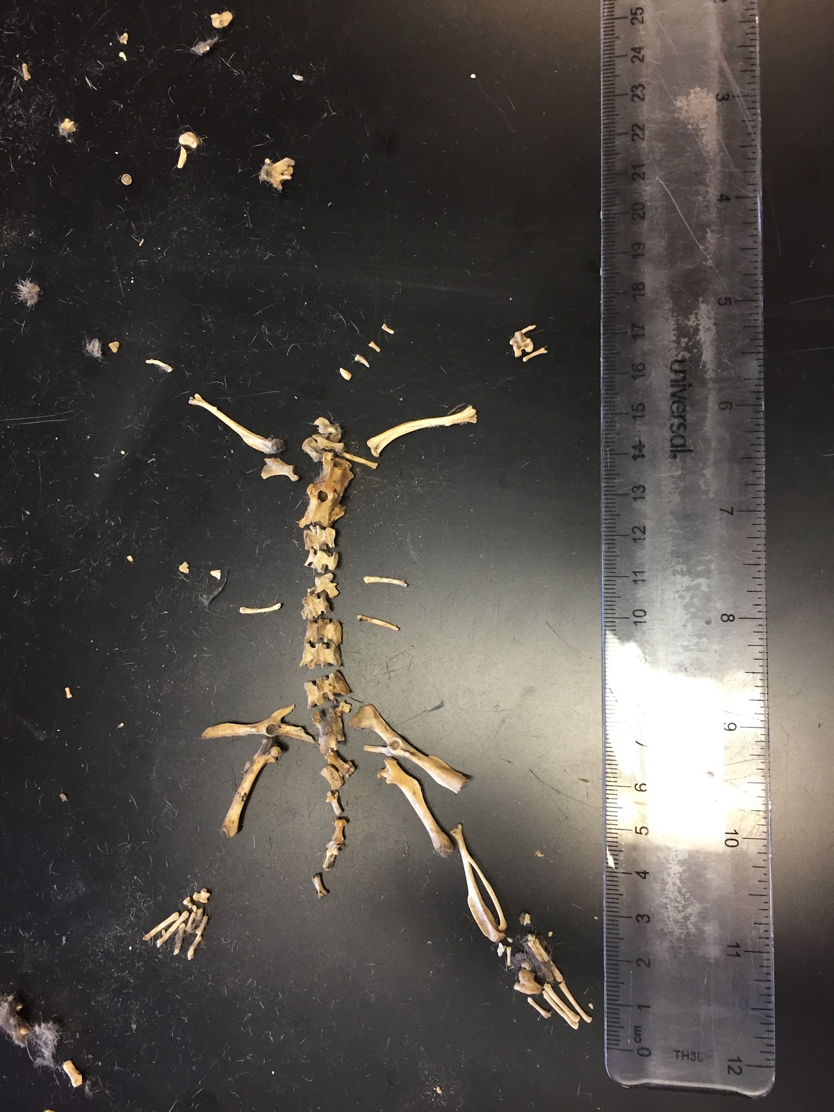
The fibula and tibia were similar to the ones on the human body because they are both connected and located in the lower leg. In both the human and vole, one bone was larger than the other; however, in the vole, the fibula was larger, while in a human the tibia is much larger. Also, the gap between the two bones on the vole was much larger and circular than it is in a human. This displays how these organisms have very similar features, but are unique in their own ways.


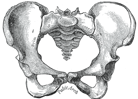
Another structure that was similar to a human's was the pelvis. Both pelvises have a gap, where the bones form loops. However, the pelvis of the vole is more triangularly shaped than the human's. The human pelvis is much wider and contains another large gap in it.

The spine of a vole was remarkably similar to the spine of a human. Both of the spines had individual pieces that connected together. Both spines appeared to have jagged edges, but the individual pieces of a human spine are much more round. The voles' spine is very jagged all around.


No comments:
Post a Comment